
TAKE ACTION
A bronchiectasis diagnosis can help you determine the right management approach for your patients1,2
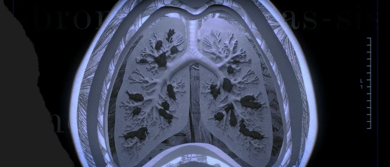
CT imaging is the standard procedure to confirm or rule out a bronchiectasis diagnosis when symptoms and clinical history suggest that it could be an underlying respiratory disease.3,4
When diagnosing bronchiectasis, a CT scan has high accuracy as well as higher sensitivity and specificity compared to an X-ray.5 Terms such as “bronchial/airway dilation,” “signet ring,” or “tree-in-bud” in a radiology report may indicate a finding of bronchiectasis on a CT scan.5-7
A bronchiectasis diagnosis can be confirmed with a CT scan and key clinical features:

Key clinical findings6
- Cough and sputum production most days of the week
- History of exacerbations

Chest CT3,4
CT imaging findings of bronchial dilation confirm a diagnosis of bronchiectasis when clinically suspected.
3 key defining radiologic features of bronchiectasis6,7
Bronchiectasis is characterized by structural lung damage that is visible on lung imaging.
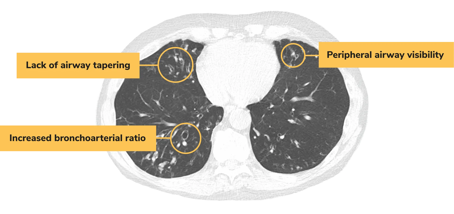
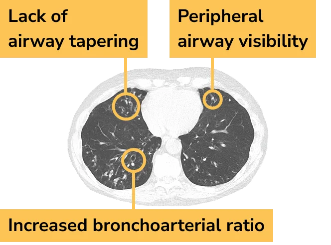
If you suspect bronchiectasis in patients with clinical markers of the disease, a CT scan in the diagnostic evaluation can confirm or rule it out.1,8
Addressing the 4 drivers of bronchiectasis may help manage its chronic and progressive nature2,9,10
Goals in the management of bronchiectasis include disrupting the vicious vortex by addressing chronic airway infection, reducing chronic airway inflammation, enhancing mucociliary clearance, and mitigating lung destruction.2,10,11
Expert commentary: Addressing the vicious vortex of bronchiectasis
Speakers: Ashwin Basavaraj, MD; Charles Daley, MD; Mark Metersky, MD; Patrick Flume, MD
All participants have been compensated for their time.
Bronchiectasis requires a multimodal treatment plan11,12
The treatment of bronchiectasis involves targeting the key components of the vicious vortex2,10,11


A different treatment approach may be required to specifically manage the underlying bronchiectasis in patients with COPD or asthma.2,4,5,11
Turn clinical suspicion into informed action.
Explore tools and resourcesCT=computed tomography.